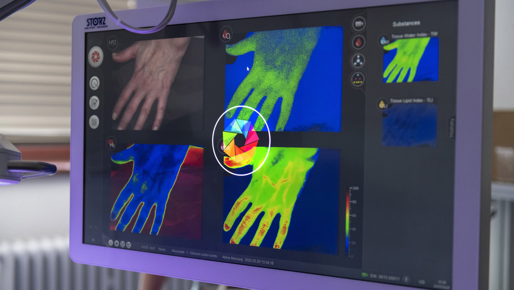
During tumor surgery, millimeters can make or break the success of the procedure. Surgeons must completely remove diseased tissue without damaging healthy structures such as nerves. A new imaging system is expected to make this work more precise in the future. As part of the STAIN collaborative project, the team led by Prof. Dr. Marianne Maktabi is developing a camera that uses invisible light and can distinguish tumor tissue from healthy tissue.
Hyperspectral Imaging as a Diagnostic Tool
The principle is well known: light is shone onto the tissue, and the reflected light is broken down into its individual wavelengths. Each tissue leaves behind a characteristic spectral signature, comparable to a fingerprint. What makes this system unique is that the intraoperative imaging system operates in the short-wavelength infrared range between 1,000 and 2,500 nanometers. In this wavelength range, additional information can be obtained about molecules in the tissue that remain hidden in other ranges.
Existing clinical procedures typically use contrast agents that are injected into the body to make tissue glow under special cameras. This fluorescence imaging carries risks: allergic reactions are possible, and not all patients tolerate the substances. Although current spectral cameras already operate without contrast agents, they only capture the visible and near-infrared spectrum up to 1,000 nanometers. Studies show that the accuracy is not yet sufficient for clinical use.
Preliminary work by the research group paves the way
The project team is building on solid preliminary work. In several scientific publications, the research group has already demonstrated that hyperspectral imaging can detect colorectal cancer and identify circulatory disorders following microsurgical tissue transplants. These findings form the basis for the next step: clinical measurements in the extended infrared range.
Two doctoral candidates, research assistants, and student assistants will work on the project. The interdisciplinary research initiative relies on a partnership with HAWK University of Applied Sciences and Arts Hildesheim/Holzminden/Göttingen, three German hospitals, and the Hanover-based company HAIP. The hospitals will provide data from the operating room and test the system under real-world conditions. HAIP will contribute technological expertise in the field of hyperspectral cameras.
Planned for Use in Two Surgical Fields
The researchers are focusing on two areas of application: In visceral surgery, the system is intended for use during bowel resections; in ear, nose, and throat (ENT) surgery, it is intended for procedures in the head and neck region. Both areas place high demands on precision because they involve many sensitive structures in a confined space.
“We are not developing a finished product here, but rather exploring potential applications,” explains project leader Prof. Dr. Marianne Maktabi, who has been working at the Department of Electrical, Mechanical and Industrial Engineering at Anhalt University of Applied Sciences since 2022. “Medical device development is complex and time-consuming. Following this project, operational clinical products can be developed in subsequent projects in collaboration with industry partners and hospitals.”
Joint Funding Through HAW-ForschungsPraxis
The Federal Ministry of Education and Research is funding STAIN with a total of 2.5 million euros under the HAW-ForschungsPraxis program. Of this amount, 1.38 million euros will go to Anhalt University of Applied Sciences, and 1.12 million euros to HAWK Göttingen. The project runs from January 1, 2026, to December 31, 2029.
“The selection process was challenging,” reports Research Focus Manager Dr. Johanna Berg from the KAT team at Anhalt University of Applied Sciences. “Of the approximately 300 project outlines submitted, only 63 were invited to submit a full proposal. The fact that our project is among the few that received funding confirms its importance for forward-looking healthcare.”
Internationally, such a system has not yet been clinically established. Although various projects are conducting research on hyperspectral imaging, the short-wavelength infrared range has not yet been comprehensively investigated. New application possibilities can be explored here.
Contribution to Digital and Preventive Medicine
The project aligns with Science Year 2026, which has the theme “Medicine of the Future.” STAIN combines two key aspects: the digital processing of information in the operating room and the prevention of complications through the early detection of risk structures.
Ultimately, many patients could benefit—not only from oncological procedures but from all surgical operations in which nerves must be preserved. The system could provide surgeons with a “technical eye” that sees more than the human eye.
Information and contact:
Prof. Dr. Marianne Maktabi
Anhalt
University of Applied Sciences Department of Electrical, Mechanical and Industrial
Engineering marianne.maktabi@hs-anhalt.de
